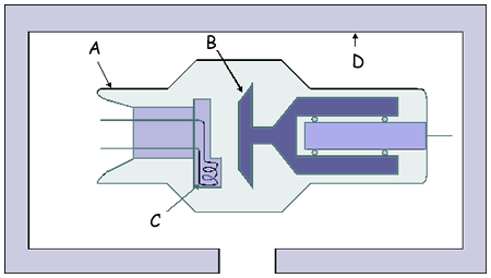
42 x ray tube labeled
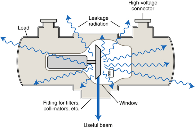
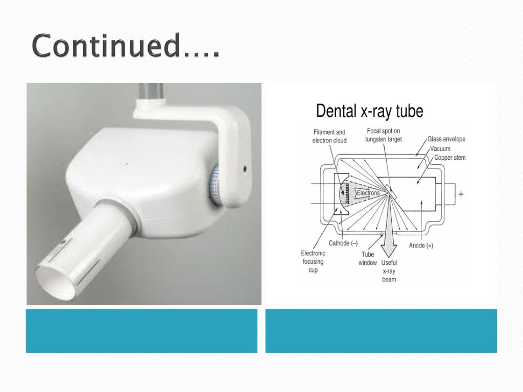
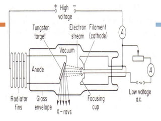
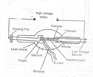
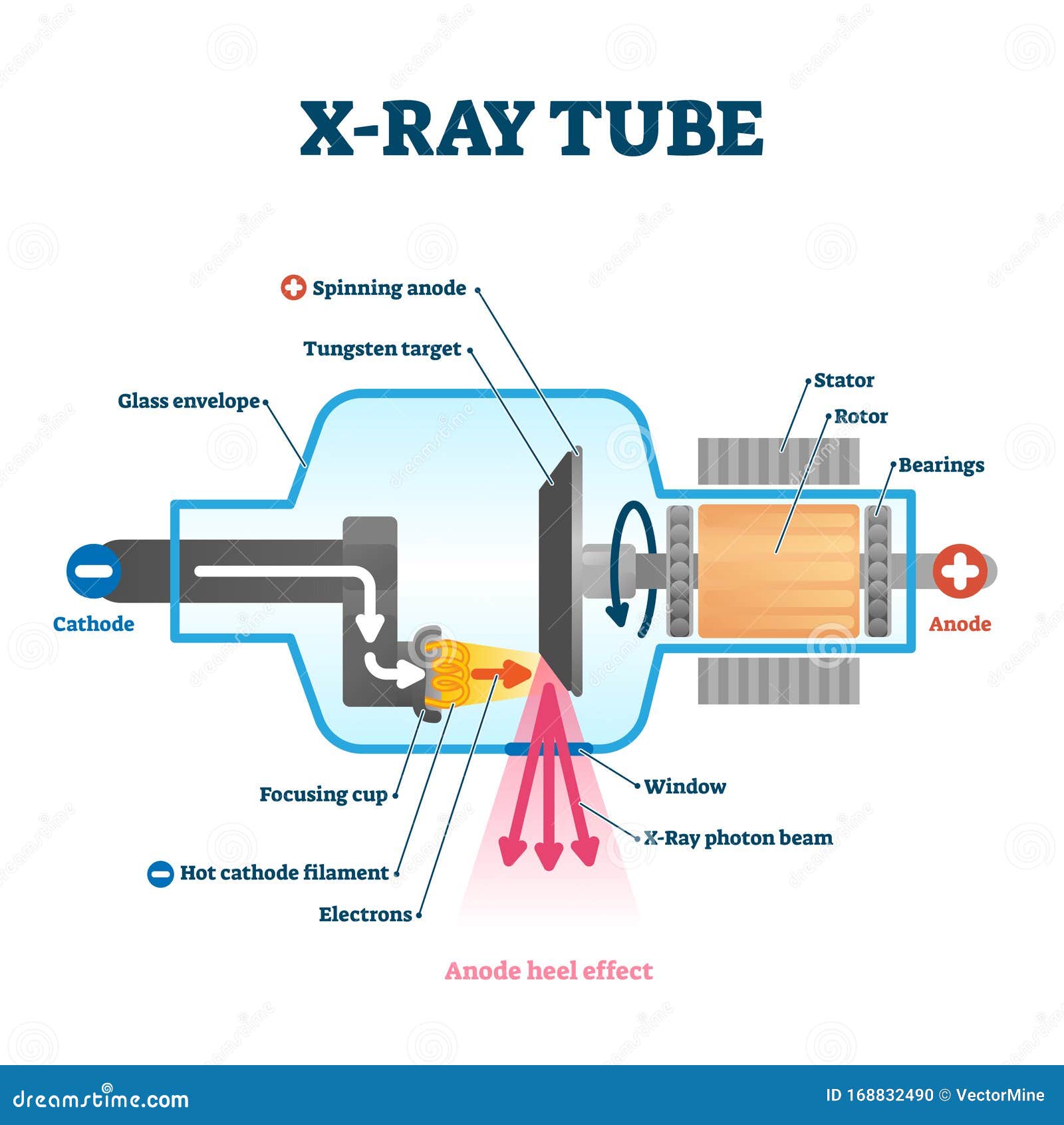
radiopaedia.org › articles › x-ray-tube-1X-ray tube | Radiology Reference Article | Radiopaedia.org Mar 11, 2022 · An x-ray tube functions as a specific energy converter, receiving electrical energy and converting it into two other forms of energy: x-radiation (1%) and heat (99%). Heat is considered the undesirable product of this conversion process; therefore x-radiation is created by taking the energy from the electrons and converting it into photons . › en › libraryRadiological anatomy: X-ray, CT, MRI | Kenhub Aug 02, 2022 · Normal chest x ray. Radiological anatomy is where your human anatomy knowledge meets clinical practice. It gathers several non-invasive methods for visualizing the inner body structures. The most frequently used imaging modalities are radiography (X-ray), computed tomography (CT) and magnetic resonance imaging (MRI). X-ray and CT require the ...
Clavicle X-ray: projections, norm, description - I Live! OK An X-ray of a healthy clavicle / X-ray of a clavicle normally gives a clear (light) image of the contour of the bone body, its ends - the sternum and shoulder, joints (acromioclavicular and sternoclavicular), as well as the humeral process of the scapula. [ 4] All structures are anatomically correct, there are no darkening. [ 5]
X ray tube labeled
unixray.com › x-ray-machine-troubleshootingThe 12 Most Common X-ray Machine Problems and Troubleshooting Firstly, there is the issue of undesirable scatter X-rays, a type of secondary radiation that results as the beam from the X-ray tube projects on the sample causing the rays to scatter. This scattering often occurs as multiple scattering but can sometimes be a single scatter X-ray beam, but both scatter X-rays result in blurry images. Gastrostomy Tube Replacement - StatPearls - NCBI Bookshelf Coagulation disorders. If the tube has been displaced for more than 24 hours, and the track has narrowed or closed. G tube (size similar to prior G-tube) 10 mL saline syringe. Guidewire. Dressing kit. 2 x 2 and 4 x 4 gauze. Alcohol swabs and povidone-iodine swabs. Gastroscope (if endoscopic replacement is being considered) X-Rays/Radiographs | American Dental Association The ADA has joined with more than 80 other health care organizations to promote Image Gently, an initiative to "child size" radiographic examination of children in medicine and dentistry. State laws and regulations set specific requirements for the use of ionizing radiation (which includes X-rays). Introduction.
X ray tube labeled. Normal chest x-ray: Anatomy tutorial | Kenhub X-ray of the chest (also known as a chest radiograph) is a commonly used imaging study, and is the most frequently performed imaging study in the United States.It is almost always the first imaging study ordered to evaluate for pathologies of the thorax, although further diagnostic imaging, laboratory tests, and additional physical examinations may be necessary to help confirm the diagnosis. Tubular shape aware data generation for segmentation in medical imaging ... Purpose Chest X-ray is one of the most widespread examinations of the human body. In interventional radiology, its use is frequently associated with the need to visualize various tube-like objects, such as puncture needles, guiding sheaths, wires, and catheters. Detection and precise localization of these tube-like objects in the X-ray images are, therefore, of utmost value, catalyzing the ... CLiP, catheter and line position dataset | Scientific Data - Nature This paper describes a dataset consisting of 50,612 image level and 17,999 manually labelled annotations from 30,083 chest radiographs from the publicly available NIH ChestXRay14 dataset with... radiopaedia.org › articles › x-ray-production-2X-ray production | Radiology Reference Article | Radiopaedia.org Nov 28, 2020 · An x-ray generator gives power to the x-ray tube. It contains high voltage transformers, filament transformers and rectifier circuits. Cathode. The cathode is the negative terminal of an x-ray tube. It is a tungsten filament and when current flows through it, the filament is heated and emits its surface electrons by a process called thermionic ...
How to check g tube placement before feeding? - Nutritionless Fill the balloon port with an empty syringe labeled "BAL". Remove the entire amount of water from the balloon. Examine what was taken away. Remove the old water and dispose of it. Using new sterile or distilled water, re-inflate the balloon. Never use saline or compressed air. When is it necessary to double-check the installation of a feeding tube? CFR - Code of Federal Regulations Title 21 - Food and Drug Administration Each such system failure override switch shall be clearly labeled as follows: For X-ray Field Limitation System Failure (c) Activation of tube. X-ray production in the fluoroscopic mode shall be... Chest Tube - StatPearls - NCBI Bookshelf The chest tube can be discontinued once no air leak is visualized, output is serosanguinous with no signs of bleeding, output is less than 150 cc to 400 cc over a 24-hour period (this range is wide because it is debatable among researchers), nonexistent or stable mild pneumothorax on chest x-ray, and the patient is minimized on positive ... Health tech company in talks with FDA about device that may have caused ... The company advertised the device as limiting the need for x-rays to verify location, but the field correction would indicate that those additional steps would in fact remain necessary for operation.
microbenotes.com › x-ray-spectroscopy-principleX-Ray Spectroscopy- Definition, Principle, Steps, Parts, Uses Jan 22, 2022 · X-rays make up X-radiation, a form of electromagnetic radiation. Most X-rays have a wavelength ranging from 0.01 to 10 nanometers, corresponding to frequencies in the range 30 petahertz to 30 exahertz (3×10¹⁶ Hz to 3×10¹⁹ Hz) and energies in the range 100 eV to 100 keV, produced by the deceleration of high-energy electrons. Nasogastric tube position on chest x-ray (summary) x-rays are only performed when the position is uncertain most tube positions are checked by assessing pH of tube aspirate normal tube descends the thorax in the midline tube bisects the carina tube crosses the diaphragm in the midline the tip sits below the diaphragm viewing the tube you need to be confident that you can see the tip X-rays | Radiology Reference Article | Radiopaedia.org X-rays (or much more rarely, and usually historically, x-radiation or Roentgen rays) represent a form of ionizing electromagnetic radiation. They are produced by an x-ray tube, using a high voltage to accelerate the electrons produced by its cathode. The produced electrons interact with the anode, thus producing x-rays. Industrial Radiography | US EPA The detector records x-rays or gamma rays that pass through the material. The thicker the material, the fewer x-rays or gamma rays can pass through. Because the material is thinner where there is a crack or flaw, more rays pass through that area. The detector creates a picture from the rays that pass through, which shows cracks or flaws.
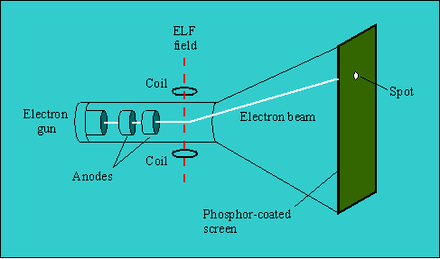
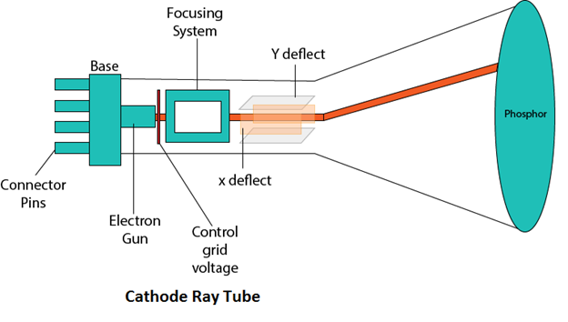
Cathode Ray Tubes (CRTs) | US EPA Cathode Ray Tubes (CRTs) A cathode ray tube (CRT) is the glass video display component of an electronic device (usually a television or computer monitor). EPA encourages repair and reuse as a responsible ways to manage CRTs. If reuse or repair are not practical options, CRTs can be recycled. Recycled CRTs are typically disassembled so that ...
X-ray artifacts | Radiology Reference Article | Radiopaedia.org black "lightning" marks resulting from films forcibly unwrapped or excessive flexing of the film crescent-shaped black lines due to fingernail pressure on the film crescent-shaped white lines due to cracked intensifying screen black film complete exposure to light. clear spots air bubbles sticking to film during processing
Chest X-ray Interpretation | A Structured Approach | Radiology | OSCE Various tubes and cables will be visible as radio-opaque lines on the chest X-ray (e.g. central line, ECG cables). Artificial heart valves. Artificial heart valves typically appear as ring-shaped structures on a chest X-ray within the region of the heart (e.g. aortic valve replacement). Pacemaker
X-ray Radiographic Patient Positioning - NCBI Bookshelf Last Update: December 15, 2021. Anterior denotes the front of a body part, while the posterior denotes the back. Superior denotes the top of a body part, while inferior denotes the bottom. Medial indicates towards the midline. Lateral indicates a location away from the midline. Proximal is towards the body's center.
CFR - Code of Federal Regulations Title 21 - Food and Drug Administration The manufacturer of the tube shall instruct the assembler who installs the new tube to attach the label to the tube housing assembly and to remove, cover, or deface the previously affixed...
CFR - Code of Federal Regulations Title 21 - Food and Drug Administration The following requirements apply when the equipment is operated on a power supply as specified by the manufacturer in accordance with the requirements of § 1020.30(h)(3) for any fixed x-ray tube potential within the range of 40 percent to 100 percent of the maximum rated. (1) Equipment having independent selection of x-ray tube current (mA).
X-Ray Generation Notes - University of Oklahoma X-Ray beams that are parallel with the narrow projection of the filament have an approximate focal shape of a square, which is usually labeled as a spot. These two focal projections are necessarily about 90 ° apart in the plane normal to the filament-anode axis.
Section R9-7-804 - Open-beam X-ray Systems, Ariz. Admin. Code § 9-7-804 ... A clearly visible warning light labeled with the words "X-RAY ON," or a similar warning located near any switch that energizes an x-ray tube, illuminated only when the tube is energized; and4. The warning devices in subsections (B)(1) through (3) shall be labeled so that their purpose is easily identified. C.
en.wikipedia.org › wiki › X-ray_tubeX-ray tube - Wikipedia An X-ray tube is a vacuum tube that converts electrical input power into X-rays. The availability of this controllable source of X-rays created the field of radiography , the imaging of partly opaque objects with penetrating radiation .
CFR - Code of Federal Regulations Title 21 - Food and Drug Administration Failure of a single component of the cabinet x-ray system shall not cause failure of both indicators to perform their intended function. One, but not both, of the indicators required by this...
X-ray film | Radiology Reference Article | Radiopaedia.org X-ray film displays the radiographic image and consists of emulsion (single or double) of silver halide (silver bromide (AgBr) is most common) which when exposed to light, produces a silver ion (Ag +) and an electron. The electrons get attached to the sensitivity specks and attract the silver ion.
en.wikipedia.org › wiki › X-ray_crystallographyX-ray crystallography - Wikipedia X-ray crystallography is the experimental science determining the atomic and molecular structure of a crystal, in which the crystalline structure causes a beam of incident X-rays to diffract into many specific directions. By measuring the angles and intensities of these diffracted beams, a crystallographer can produce a three-dimensional picture of the density of electrons within the crystal.
CFR - Code of Federal Regulations Title 21 - Food and Drug Administration The manufacturer shall permanently affix or inscribe a warning label, clearly legible under conditions of service, on all television receivers which could produce radiation exposure rates in excess...
Abdominal X-ray Interpretation (AXR) | Radiology - Geeky Medics Bony structures commonly visible on abdominal X-ray include: Ribs Lumbar vertebrae Sacrum Coccyx Pelvis Proximal femurs A wide range of bony pathologies can be identified on abdominal X-ray including fractures, osteoarthritis, Paget's disease and bony metastases. Calcification and artefact
X-Rays/Radiographs | American Dental Association The ADA has joined with more than 80 other health care organizations to promote Image Gently, an initiative to "child size" radiographic examination of children in medicine and dentistry. State laws and regulations set specific requirements for the use of ionizing radiation (which includes X-rays). Introduction.
Gastrostomy Tube Replacement - StatPearls - NCBI Bookshelf Coagulation disorders. If the tube has been displaced for more than 24 hours, and the track has narrowed or closed. G tube (size similar to prior G-tube) 10 mL saline syringe. Guidewire. Dressing kit. 2 x 2 and 4 x 4 gauze. Alcohol swabs and povidone-iodine swabs. Gastroscope (if endoscopic replacement is being considered)
unixray.com › x-ray-machine-troubleshootingThe 12 Most Common X-ray Machine Problems and Troubleshooting Firstly, there is the issue of undesirable scatter X-rays, a type of secondary radiation that results as the beam from the X-ray tube projects on the sample causing the rays to scatter. This scattering often occurs as multiple scattering but can sometimes be a single scatter X-ray beam, but both scatter X-rays result in blurry images.












Post a Comment for "42 x ray tube labeled"