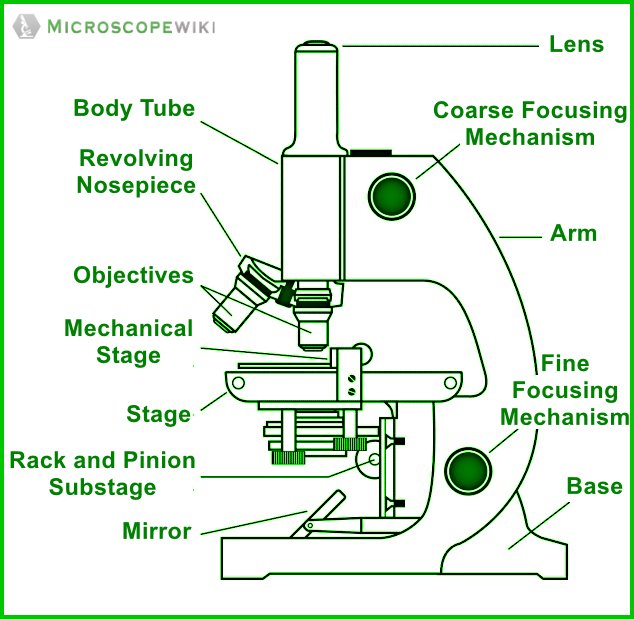
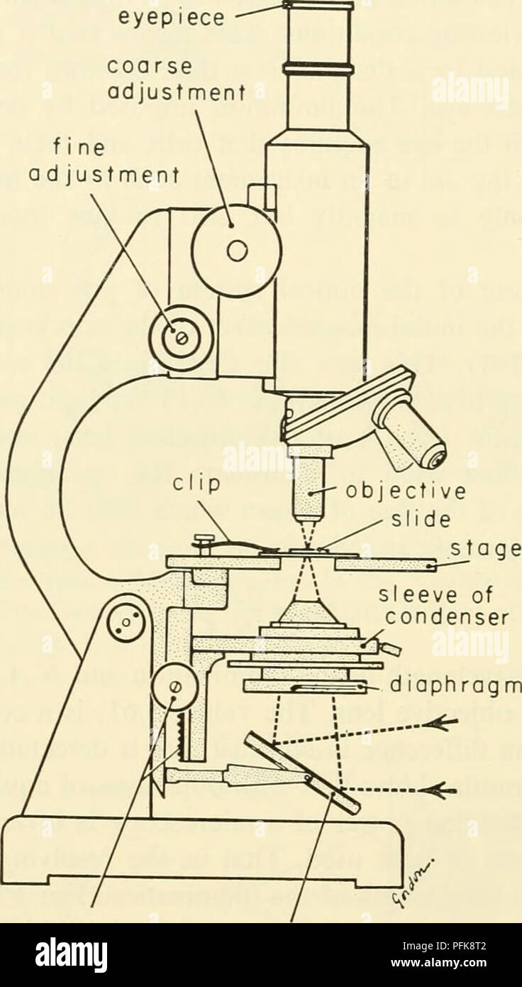
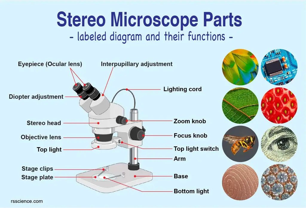
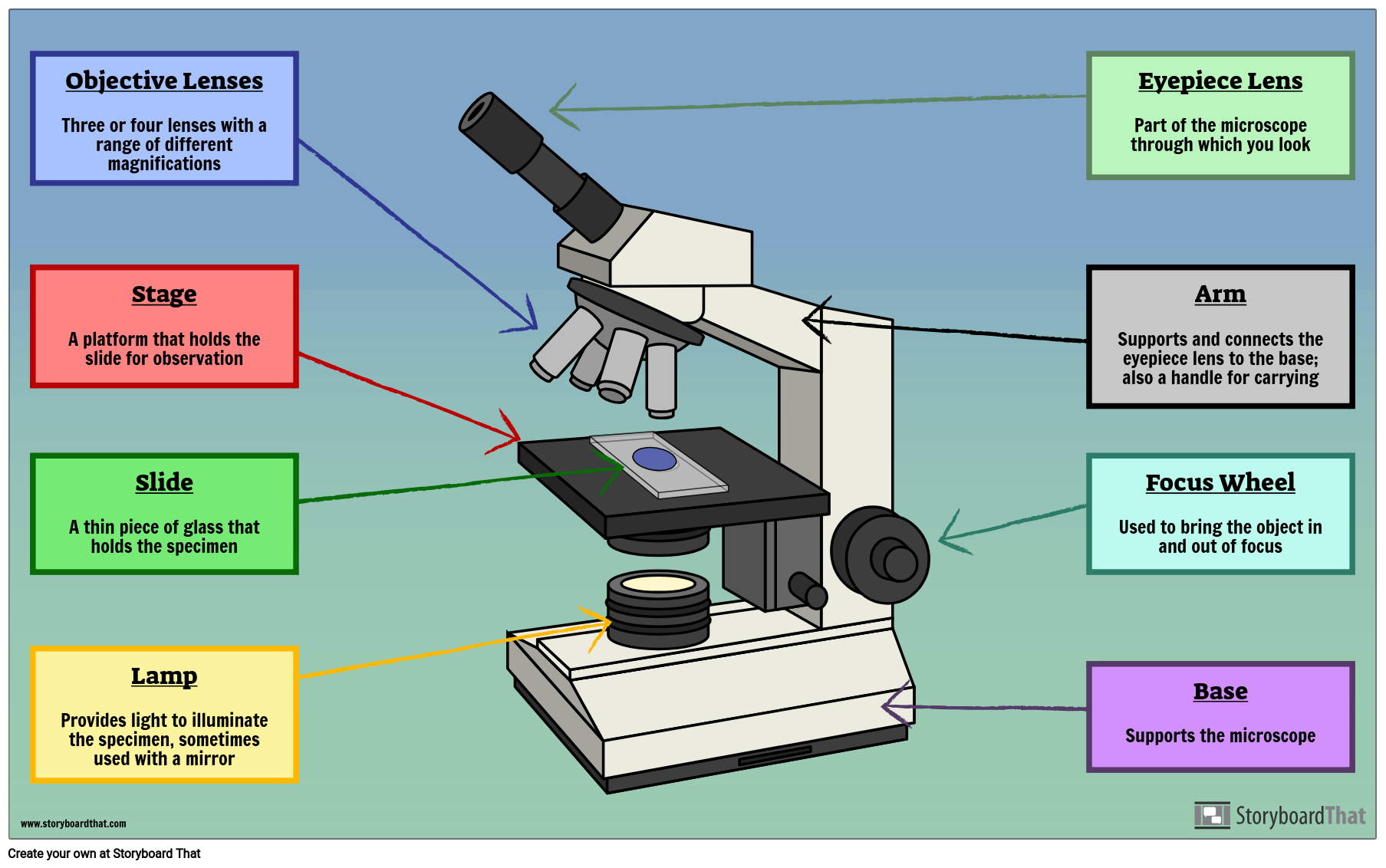
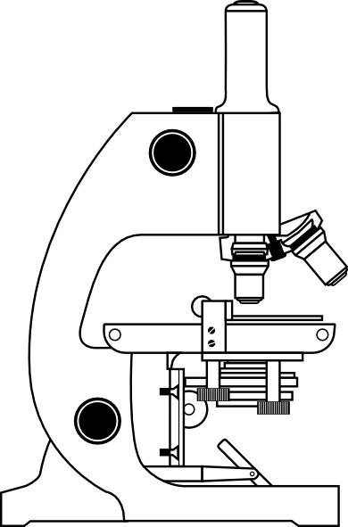
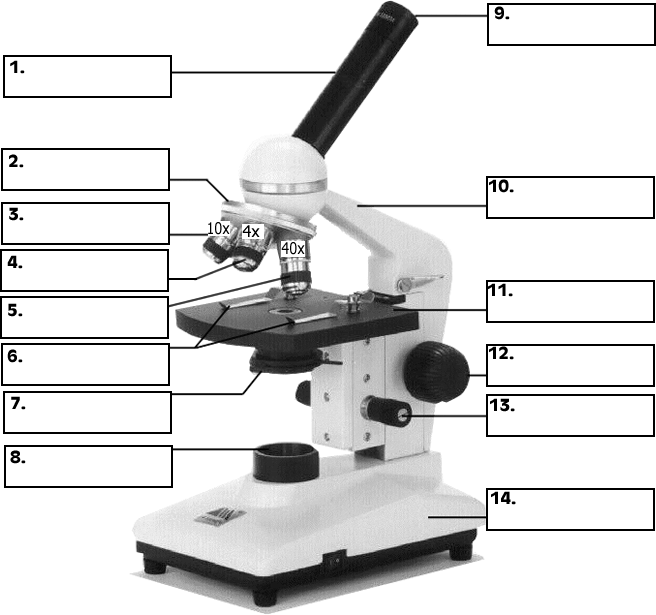
45 microscope drawing labeled
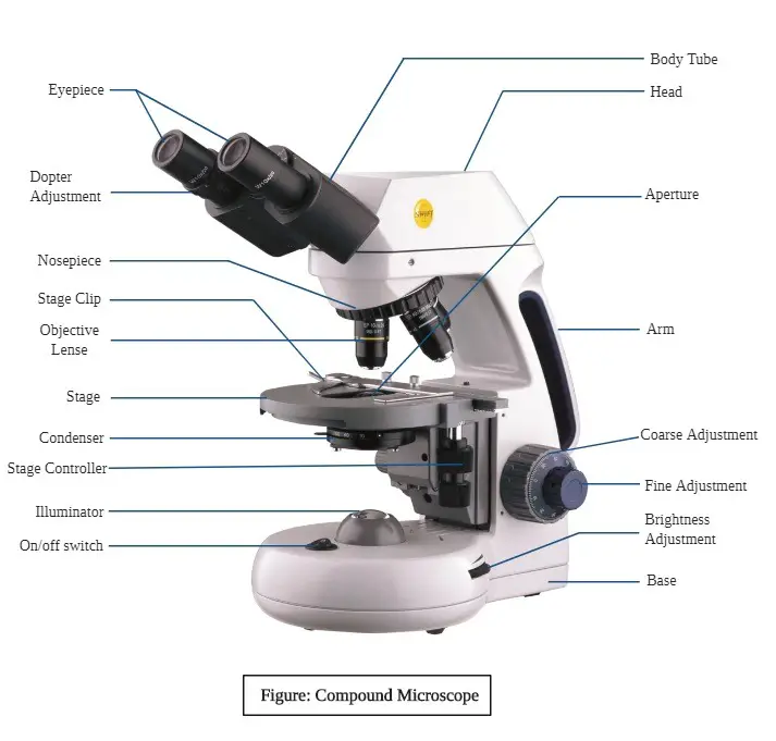
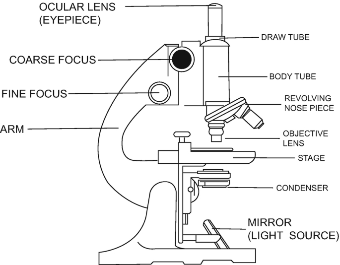
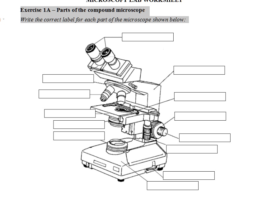
Microscope Types (with labeled diagrams) and Functions Compound microscope labeled diagram Compound microscope functions: It finds great application in areas of pathology, pedology, forensics etc Its greater order of magnification allows for deeper study of microbial organisms to Detect the cause of diseases Study the mineral composition in soils Best 10 Microscope in Lithonia, GA - YP.com Microscope in Lithonia on YP.com. See reviews, photos, directions, phone numbers and more for the best Microscopes in Lithonia, GA.
Welcome to Butler County Recorders Office Copy and paste this code into your website. Your Link …
Microscope drawing labeled
Southern Microscope in Lithonia, GA with Reviews - YP.com Find 2 listings related to Southern Microscope in Lithonia on YP.com. See reviews, photos, directions, phone numbers and more for Southern Microscope locations in Lithonia, GA. Compound Microscope Parts - Labeled Diagram and their Functions Labeled diagram of a compound microscope Major structural parts of a compound microscope There are three major structural parts of a compound microscope. The head includes the upper part of the microscope, which houses the most critical optical components, and the eyepiece tube of the microscope. Private Label 8200 Mall Pkwy Lithonia, GA - MapQuest Private Label 8200 Mall Pkwy Lithonia GA 30038 (404) 850-9552 Website. Menu & Reservations Make Reservations . Order Online Tickets Tickets See Availability Directions {{::location.tagLine.value.text}} Sponsored Topics. Legal. Help Get directions, reviews and information for Private Label in Lithonia, GA. ...
Microscope drawing labeled. Testis Histology – Complete Guide to Learn ... - AnatomyLearner Mar 01, 2021 · Learn testis histology side from labeled diagram online. This is the best guide to learn testis histology with anatomy learner ... You will also get testis histology drawing at the end of this ... I would like to enlist the structures that you should identify under the light microscope from testis histology slide at laboratory. #1. Tunica vagzi ... Labeled Microscope Diagram Clipart Free Download 291 Labeled Microscope Diagram clipart free images in AI, SVG, EPS or CDR. Save 15% on iStock using the promo code. CLIPARTLOGO15 apply promocode. Diagram Vector 3. Realistic brand cosmetic bottles. Minimalist labeled black containers design, beauty products packages, pumps and spray mockups. Vector set. Microscope Diagram Labeled Clipart Free Download 291 Microscope Diagram Labeled clipart free images in AI, SVG, EPS or CDR. Save 15% on iStock using the promo code. CLIPARTLOGO15 apply promocode. Flower type of line drawing vector diagram-6. Cells of living things. Biology and root of the plant under a microscope. Cells - University of Utah Virtual Microscope. Learn how cells work together in tissues, organs, and organ systems. Cells in Perspective. In 1665, Robert Hooke coined the term cell to describe the structures he could see in cork with some of the first microscopes. Since then, technology has given us an increasingly complex view of the basic unit of life.
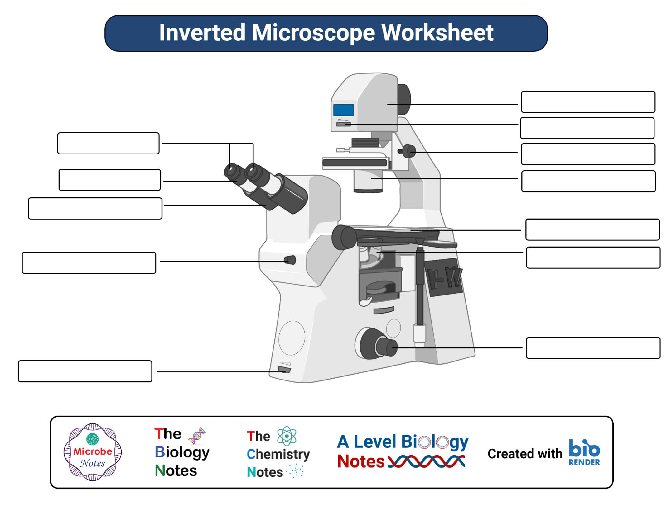
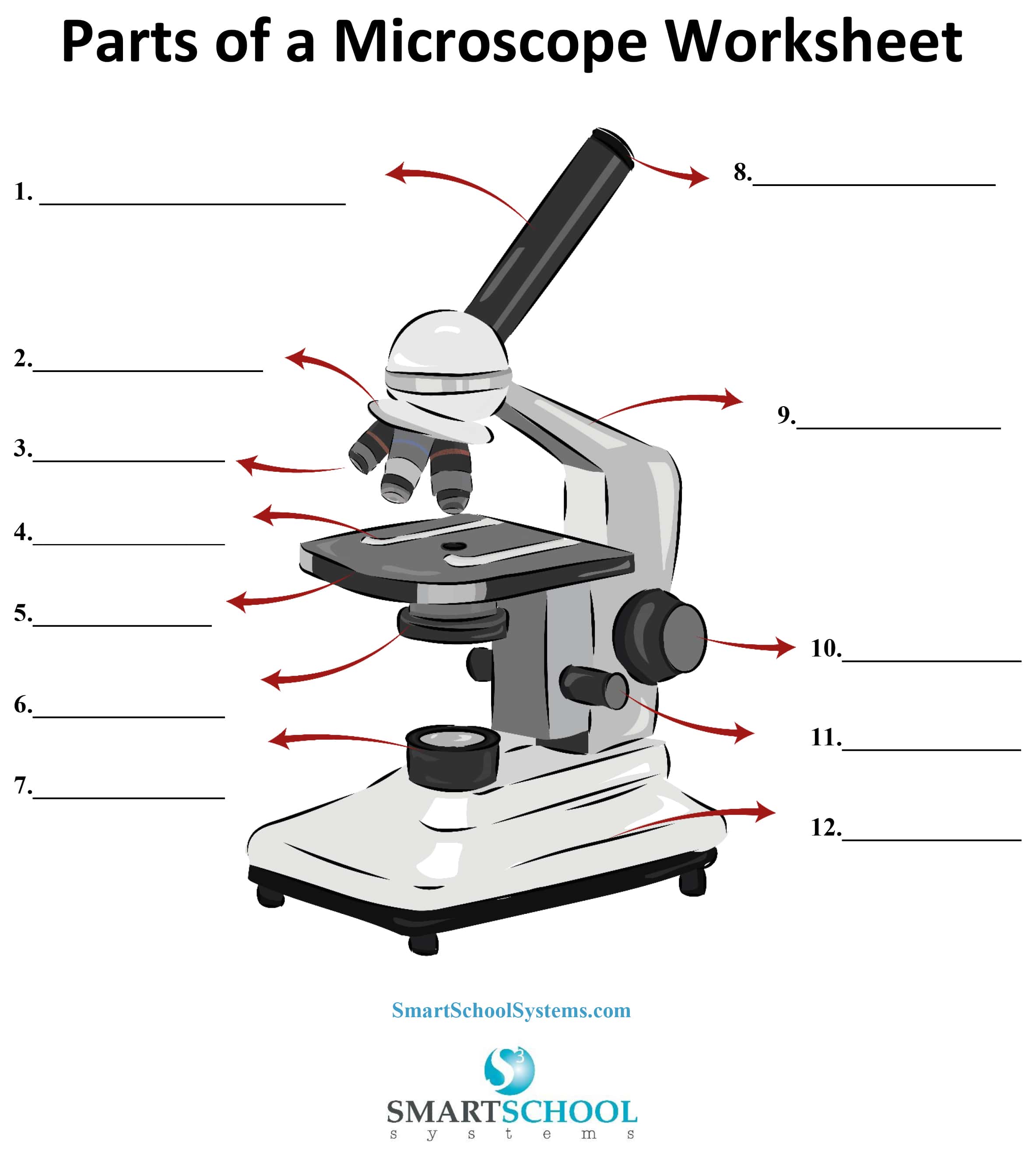
en.wikipedia.org › wiki › MicroscopyMicroscopy - Wikipedia The field of microscopy (optical microscopy) dates back to at least the 17th-century.Earlier microscopes, single lens magnifying glasses with limited magnification, date at least as far back as the wide spread use of lenses in eyeglasses in the 13th century but more advanced compound microscopes first appeared in Europe around 1620 The earliest practitioners of microscopy include Galileo ... Drawing Of Microscope And Label - Warehouse of Ideas Microscope Diagram Labeled, Unlabeled and Blank Parts of from . How to sketch a microscope slide will feel less overwhelming breaking down the drawing process. We have an article covering the history, types, and evolution of all kinds of microscopes. Yet even with the technology to digital capture images, many scientists still depend on their ... Microscope Drawing Easy with Label - YouTube Microscope Drawing Easy with Label 886 views Apr 13, 2020 In this video I go over a microscope drawing that is easy with label. There is a blank copy at the end of the video to review on your own.... Label the microscope — Science Learning Hub In this interactive, you can label the different parts of a microscope. Use this with the Microscope parts activity to help students identify and label the main parts of a microscope and then describe their functions. Drag and drop the text labels onto the microscope diagram.
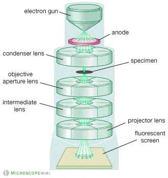
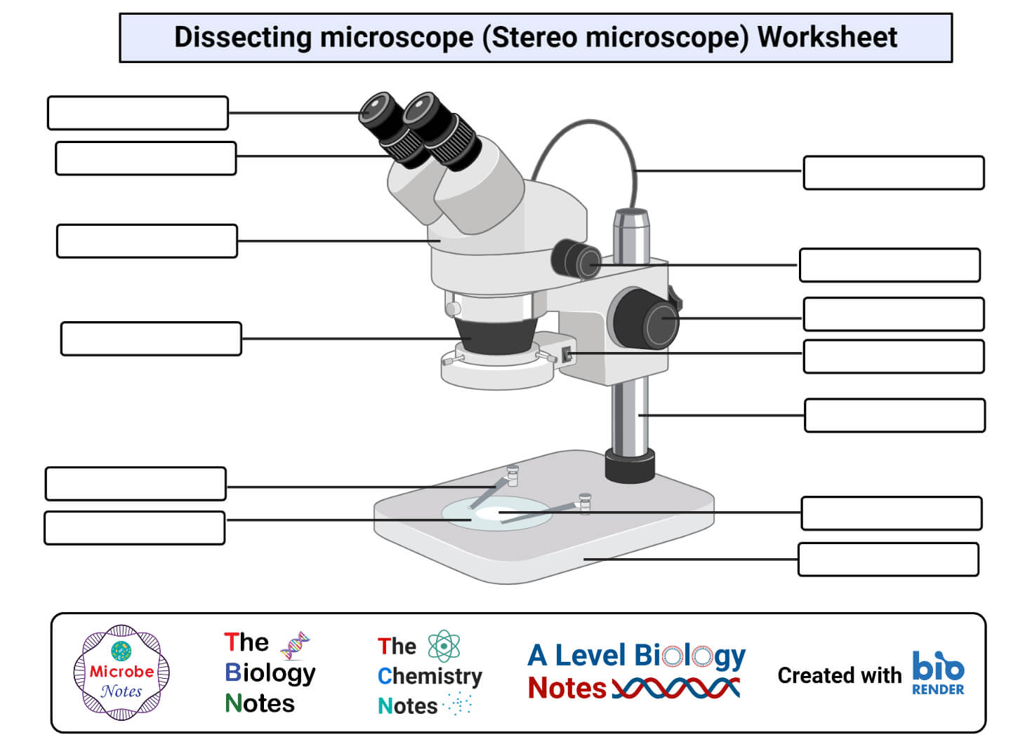
Microscopy - Wikipedia Microscopy is the technical field of using microscopes to view objects and areas of objects that cannot be seen with the naked eye (objects that are not within the resolution range of the normal eye). There are three well-known branches of microscopy: optical, electron, and scanning probe microscopy, along with the emerging field of X-ray microscopy. [citation needed] › newgrouppage9Reflective Microscope Objectives - Thorlabs Apr 12, 2022 · If using a 20X Nikon objective and Nikon trinoculars with 10X eyepieces, then the image at the eyepieces has 20X × 10X = 200X magnification. Note that the image at the eyepieces does not pass through the camera tube, as shown by the drawing to the right. Using an Objective with a Microscope from a Different Manufacturer Parts of Stereo Microscope (Dissecting microscope) - labeled diagram ... Labeled part diagram of a stereo microscope Major structural parts of a stereo microscope There are three major structural parts of a stereo microscope. The viewing Head includes the upper part of the microscope, which houses the most critical optical components, including the eyepiece, objective lens, and light source of the microscope. UD Virtual Compound Microscope - University of Delaware ©University of Delaware. This work is licensed under a Creative Commons Attribution-NonCommercial-NoDerivs 2.5 License.Creative Commons Attribution-NonCommercial-NoDerivs 2.5 …
Microscope, Microscope Parts, Labeled Diagram, and Functions Multiply the magnification of the eyepiece (ocular lens) by the magnification of the objective lens in use to calculate the total magnification of any object viewed under the microscope. This can be demonstrated using the formula. Total magnification = ocular lens x objective lens
Wikipedia:Citation needed - Wikipedia To ensure that all Wikipedia content is verifiable, Wikipedia provides a means for anyone to question an uncited claim.If your work has been tagged, please provide a reliable source for the statement, and discuss if needed.. You can add a citation by selecting from the drop-down menu at the top of the editing box.In markup, you can add a citation manually using ref tags.
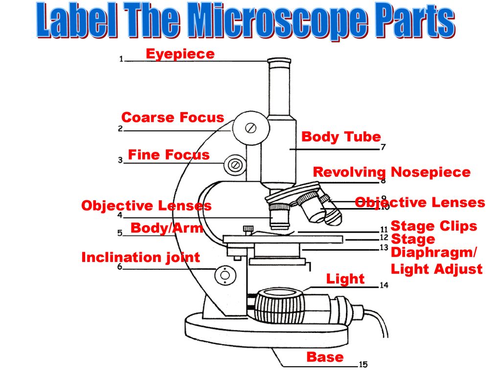
Microscope Parts and Functions Microscope Parts and Functions With Labeled Diagram and Functions How does a Compound Microscope Work?. Before exploring microscope parts and functions, you should probably understand that the compound light microscope is more complicated than just a microscope with more than one lens.. First, the purpose of a microscope is to magnify a small object or to magnify the fine details of a larger ...
www1.udel.edu › biology › ketchamUD Virtual Compound Microscope - University of Delaware ©University of Delaware. This work is licensed under a Creative Commons Attribution-NonCommercial-NoDerivs 2.5 License.Creative Commons Attribution-NonCommercial-NoDerivs 2
anatomylearner.com › testis-histologyTestis Histology - Complete Guide to Learn Histological ... Mar 01, 2021 · Testis histology labeled diagram is so important to understand the all structures of testis. In this article I am going to discuss on testis histology of animal. You will get an ideal concept of seminiferous tubule histology with a labeled diagram. I am going to share the real testis histology labeled slide with you.
Compound Microscope- Definition, Labeled Diagram, Principle, … Apr 03, 2022 · A compound microscope is of great use in pathology labs so as to identify diseases. Various crime cases are detected and solved by drawing out human cells and examining them under the microscope in forensic laboratories. The presence or absence of minerals and the presence of metals can be identified using compound microscopes.
EOF
Parts of a microscope with functions and labeled diagram - Microbe Notes Parts of a microscope with functions and labeled diagram April 19, 2022 by Faith Mokobi Having been constructed in the 16th Century, Microscopes have revolutionalized science with their ability to magnify small objects such as microbial cells, producing images with definitive structures that are identifiable and characterizable.
micro.magnet.fsu.eduMolecular Expressions: Images from the Microscope May 19, 2020 · Fluorescence Microscope Light Pathways - This interactive tutorial explores illumination pathways in the Olympus BX51 research-level upright microscope. The microscope drawing presented in the tutorial illustrates a cut-away diagram of the Olympus BX51 microscope equipped with a vertical illuminator and lamphouses for both diascopic (tungsten ...
microbenotes.com › compound-microscope-principleCompound Microscope- Definition, Labeled Diagram, Principle ... Apr 03, 2022 · A compound microscope is of great use in pathology labs so as to identify diseases. Various crime cases are detected and solved by drawing out human cells and examining them under the microscope in forensic laboratories. The presence or absence of minerals and the presence of metals can be identified using compound microscopes.
Simple Microscope - Diagram (Parts labelled), Principle, Formula and Uses A simple microscope consists of Optical parts Mechanical parts Labeled Diagram of simple microscope parts Optical parts The optical parts of a simple microscope include Lens Mirror Eyepiece Lens A simple microscope uses biconvex lens to magnify the image of a specimen under focus.
Microscope Jobs, Employment in Lithonia, GA | Indeed.com 38 Microscope jobs available in Lithonia, GA on Indeed.com. Apply to Analyst, Research Specialist, Participant and more!
en.wikipedia.org › wiki › Wikipedia:Citation_neededWikipedia:Citation needed - Wikipedia If someone tagged your contributions with a "Citation needed" tag or tags, and you disagree, discuss the matter on the article's talk page.The most constructive thing to do in most cases is probably to supply the reference(s) requested, even if you feel the tags are "overdone" or unnecessary.
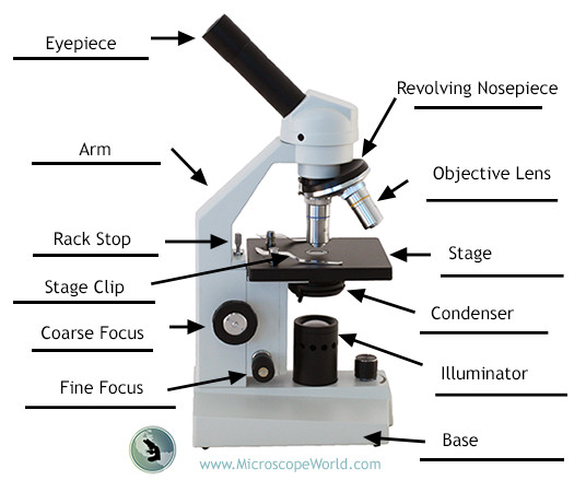
Compound Microscope Parts, Functions, and Labeled Diagram Compound Microscope Parts, Functions, and Labeled Diagram Parts of a Compound Microscope Each part of the compound microscope serves its own unique function, with each being important to the function of the scope as a whole.
American Family News Aug 02, 2022 · American Family News (formerly One News Now) offers news on current events from an evangelical Christian perspective. Our experienced journalists want to …
Reflective Microscope Objectives - Thorlabs Apr 12, 2022 · If using a 20X Nikon objective and Nikon trinoculars with 10X eyepieces, then the image at the eyepieces has 20X × 10X = 200X magnification. Note that the image at the eyepieces does not pass through the camera tube, as shown by the drawing to the right. Using an Objective with a Microscope from a Different Manufacturer
Labelled Diagram of Compound Microscope The below mentioned article provides a labelled diagram of compound microscope. Part # 1. The Stand: The stand is made up of a heavy foot which carries a curved inclinable limb or arm bearing the body tube. The foot is generally horse shoe-shaped structure (Fig. 2) which rests on table top or any other surface on which the microscope in kept.
Molecular Expressions: Images from the Microscope May 19, 2020 · Fluorescence Microscope Light Pathways - This interactive tutorial explores illumination pathways in the Olympus BX51 research-level upright microscope. The microscope drawing presented in the tutorial illustrates a cut-away diagram of the Olympus BX51 microscope equipped with a vertical illuminator and lamphouses for both diascopic (tungsten ...
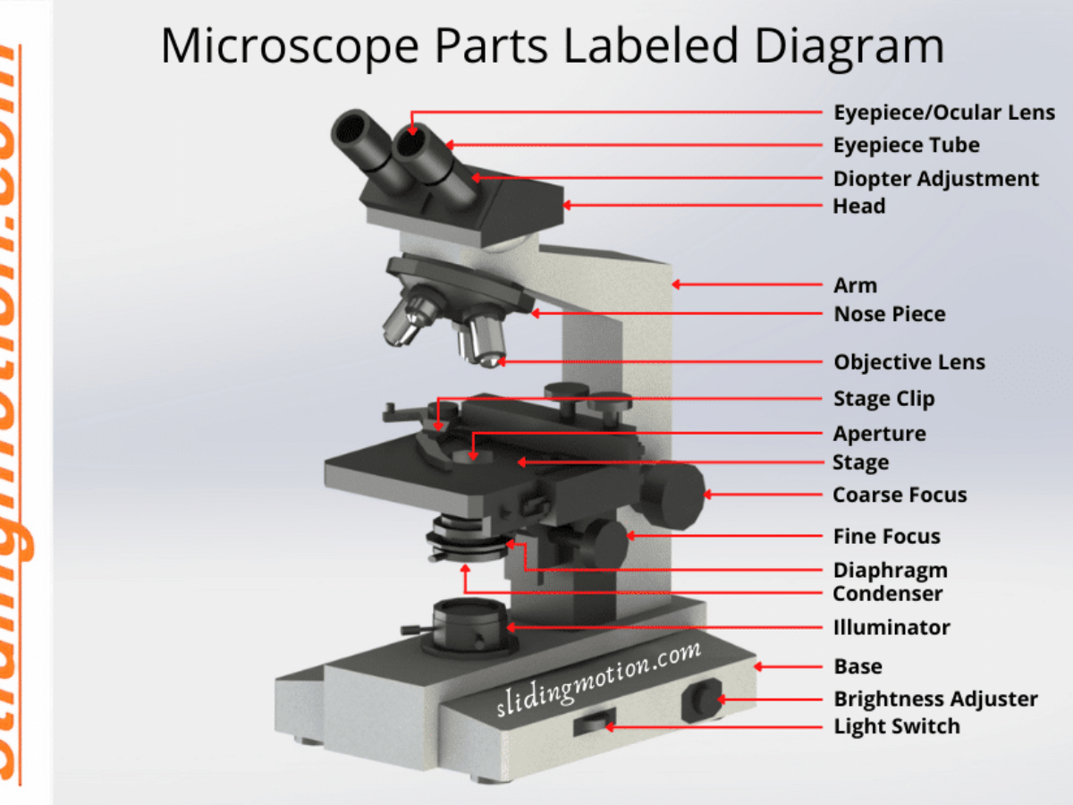
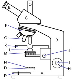
Microscope Parts, Function, & Labeled Diagram - slidingmotion Microscope parts labeled diagram gives us all the information about its parts and their position in the microscope. Microscope Parts Labeled Diagram The principle of the Microscope gives you an exact reason to use it. It works on the 3 principles. Magnification Resolving Power Numerical Aperture. Parts of Microscope Head Base Arm Eyepiece Lens
Private Label 8200 Mall Pkwy Lithonia, GA - MapQuest Private Label 8200 Mall Pkwy Lithonia GA 30038 (404) 850-9552 Website. Menu & Reservations Make Reservations . Order Online Tickets Tickets See Availability Directions {{::location.tagLine.value.text}} Sponsored Topics. Legal. Help Get directions, reviews and information for Private Label in Lithonia, GA. ...
Compound Microscope Parts - Labeled Diagram and their Functions Labeled diagram of a compound microscope Major structural parts of a compound microscope There are three major structural parts of a compound microscope. The head includes the upper part of the microscope, which houses the most critical optical components, and the eyepiece tube of the microscope.
Southern Microscope in Lithonia, GA with Reviews - YP.com Find 2 listings related to Southern Microscope in Lithonia on YP.com. See reviews, photos, directions, phone numbers and more for Southern Microscope locations in Lithonia, GA.













Post a Comment for "45 microscope drawing labeled"